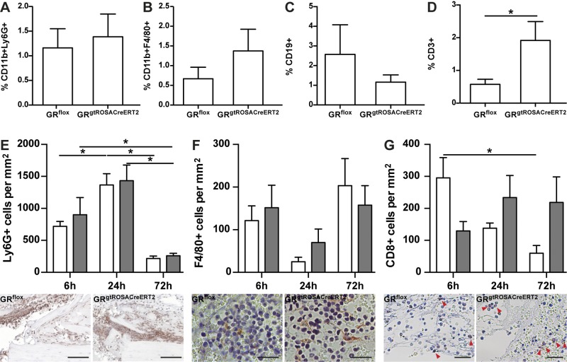
Figure 2.
Deletion of GR moderately influences inflammatory phase after fracture. Immune-cell populations infiltrating hematoma, including CD11b+Ly6G+ (PMNs, A), CD11b+F4/80+ (monocyte/macrophage lineage, B) CD19+ cells (B cells; C), and CD3+ cells (T cells; D), were analyzed by flow cytometry. Immunohistochemical staining of Ly6G (E), F4/80 (F), and CD8 (G) was performed to confirm flow cytometry results. White bars, GRflox mice; gray bars, GRgtROSACreERT2 mice. Micrographs directly below respective diagrams depict respective immune-cell population 24 h after osteotomy. Scale bars, 50 µm (E); 25 µm (F, G). Data are presented as means ± sem. Flow cytometry n = 5–6 per genotype, immunohistochemistry n = 3–6 per group. *P ≤ 0.05.

