Figure 6.
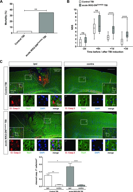
Acute neuronal IKK2-DN transgene expression also results in detrimental TBI outcome. A) Mortality of mice with acute neuronal NF-κB inhibition after CHI. The mortality rate is depicted as the percentage of animals that died. Acute IKK2-DNCamk2a mice showed a significantly enhanced posttraumatic mortality rate compared to control littermates, similar to the chronic IKK2-DNCamk2a mouse model. (Control TBI, n = 30; Acute IKK2-DNCamk2a TBI, n = 17). **P < 0.01, vs. control, by Fisher’s exact test. B) NSS score of mice with acute neuronal NF-κB inhibition. Head-injured acute IKK2-DNCamk2a mice also showed a delayed recovery from TBI, as reflected by a significantly increased NSS, compared to head-injured control animals. Data are presented as box plots with median ± interquartile range; whiskers show minimum and maximum range (n = 11–22). ****P < 0.0001 [not significant (ns), by nonparametric Mann-Whitney U test]. C) Expression of apoptotic cells in mice with acute neuronal NF-κB repression. Increased posttraumatic neuronal cell death 3 d after TBI in the injured (ipsi) hemisphere of acute IKK2-DNCamk2a mice vs. control animals and the uninjured (contra) hemisphere. Immunofluorescent staining of cleaved caspase 3+ neurons (TUJ1+ cells). Quantification of cleaved caspase 3+ cells indicated significantly enhanced apoptosis in mice with acute neuronal NF-κB inhibition vs. control littermates. Means ± sem (n = 3–4). *P < 0.05, **P < 0.01, ****P < 0.0001 (by 1-way ANOVA with Bonferroni’s correction). Scale bars, 500 µm; i′, inset′: 100 µm; i″, inset″: 20 µm.

