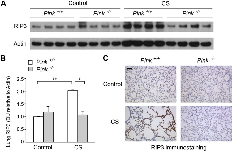
Figure 5.
Effect of PINK1 on lung necroptosis marker RIP3 during chronic CS exposure. A) Immunoblot of RIP3 and β-actin (as loading control) in wild-type (Pink1+/+) and Pink1−/− mouse lung homogenates following exposure to ambient air control or CS (6 mo). B) Bar graphs of RIP3 protein abundance in mouse lungs measured by densitometry; means ± sem. DU, densitometry units. *P < 0.05, **P < 0.01 (n = 4). C) Representative immunohistochemistry image of RIP3 in lung parenchyma of Pink1+/+ and Pink1−/− mice chronically exposed to CS (6 mo). Scale bar, 50 μm. Image is representative of 5 images/mouse lung; n = 3 mice/group.

