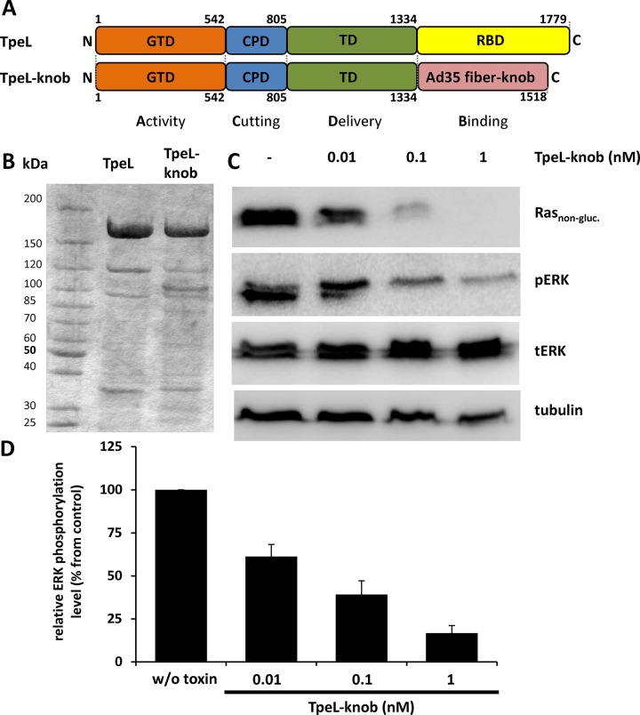
Figure 6. TpeLGTD is targeted by the fiber knob to CD46-positive cell line Capan-2.
(A) Schematic representation of the domain architecture of TpeL in comparison to the newly created fusion protein TpeL-knob. GTD, glycosyltransferase domain; CPD, cysteine protease domain; TD, translocation domain; RBD, receptor binding domain. (B) Coomassie-stained SDS-PAGE gel comparing the purity of TpeL and TpeL-knob proteins used in this study. (C) Western blot of Capan-2 cell lysates probed for Rasnon-gluc., pERK, tERK, and tubulin. Prior to lysis cells were treated with increasing concentrations of TpeL-knob for 16 h. A representative blot of three independent experiments is shown. (D) Statistical analysis of the amount of phosphorylated ERK following toxin treatment as presented in C.

