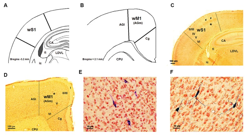
Fig.1.

wS1/M1cortical areas and NADPH-d labeled neurons. A, B. Schematic representation sections of the wS1 and wM1 cortices, respectively, modified from the Paxinos and Watson atlas, C, D. Histograms showing coronal sections of the wS1 and wM1, respectively, E, and F. Highmagnification of some NADPH-d labeled neurons in layer V of the wS1 and wM1 cortices which is related to the asterisks places in part B and E, respectively. AGl; Agranular lateral field, AGm; Agranular medial field, CA1; Corn of amons of hippocampus, Cg; Cingulate area, CPU; Caudate putamen, ic; Internal capsule, fi; Fimberia, LDVL; Laterodorsal thalamic nucleus, ventrolateral part, wS1 and wM1; Whisker part of somatosensory and primarymotor cortices.
