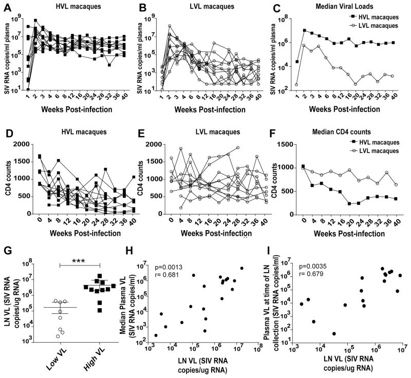
Figure 1.
Virological and immunological status of the study animals. (A–B) Viral load of HVL and LVL animals, respectively, over the course of infection. (C) Median viral load of the HVL and LVL groups post-infection. (D–E) CD4 counts of HVL and LVL animals, respectively, post infection. (F) Median CD4 counts of the HVL and LVL groups post-infection. (G) LN viral loads of the macaques. LN cells from 8 LVL and 11 HVL macaques were available for analysis. (H) Correlation between LN viral loads and median plasma viral load. (I) Correlation between LN viral loads and plasma viral loads at the time of LN collection. Viral loads at time of LN collection for 2 HVL macaques were not determined. Data of panel G were analyzed by the Mann-Whitney test. Data of panels H–I were analyzed by the Spearman correlation test. * * * P < 0.001.

