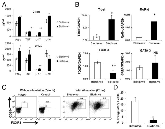
FIGURE 1.
Biotin deficiency enhances proinflammatory cytokine secretion in anti-CD3/CD28–stimulated CD4+ T cells. CD4+ T cells were stimulated with CD3/CD28 magnetic beads and cultured in biotin-deficient and -sufficient AIM V medium for 24 and 72 h. (A) Bar graph depicts the levels of IFN-γ, TNF, IL-17, and IL-10 in the supernatants of CD4+ T cells cultured in biotin+ve (sufficient) and biotin−ve (deficient) media. (B) Bar graphs depict the levels of TFs T-bet, RORγt, Foxp3, and GATA-3 in the cells, as determined by quantitative real-time PCR. GAPDH was used as control. (C) Dot plots depict the percentages of CD4+CD25+Foxp3+ Tregs in unstimulated cells and cells stimulated for 72 h. (D) Bar graph depicts the cumulative percentage of Tregs. Data are mean ± SE of six experiments with different donors. *p ≤ 0.05, **p ≤ 0.01.

