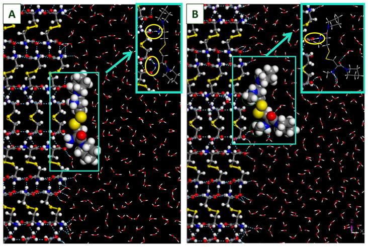
Figure 4.
Structure configurations of CDPIP (LH706, 2f) and Me2-CDPIP (LH714, 3f), respectively, adsorbed onto the {100} surface of L-cystine crystal (in ball-n-stick representation). CDPIP and Me2-CDPIP molecules are in CPK representation at 70% of vdW radii; solvent (H2O) molecules are in line representation. The methyl groups in Me2-CDPIP are circled in green. Insets are made to show hydrogen bonding details (circled in yellow), with the CDPIP and Me2-CDPIP shown in line representation). Red: oxygen; grey: carbon; white: hydrogen; blue: nitrogen; yellow: sulfur.

