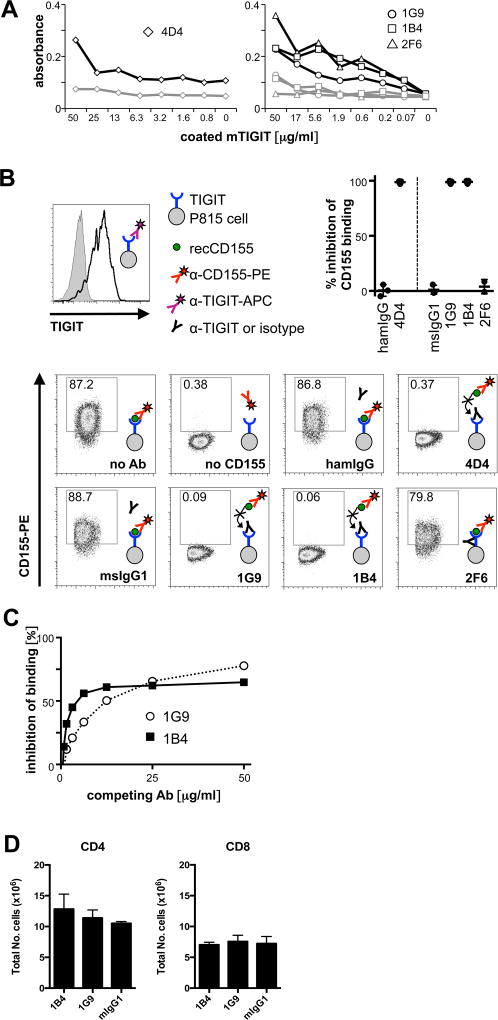
Figure 1. Anti-TIGIT antibodies compete with CD155.
TIGIT-specific antibodies were generated in armenian hamster (clone 4D4) or TIGIT−/− mice (clones 1G9, 1B4, and 2F6). (A) anti-TIGIT antibodies were titrated in an ELISA against recombinant mouse TIGIT (black) or a control (grey) protein. (B) P815 cells transfected with mouse TIGIT that uniformly express TIGIT (top left) were incubated with anti-TIGIT or isotype control antibody (Armenian hamster IgG for clone 4D4 or mouse IgG1 for 1G9, 1B4, and 2F6). Samples were then incubated with recombinant mouse CD155, then stained with anti-CD155 and analyzed by flow cytometry. A sample not incubated with CD155 was used as a positive control for blocking of CD155 binding. Representative FACS plots and summary data of inhibition of CD155 binding of 3–5 independent experiments are shown. (C) TIGIT-expressing splenocytes from TIGIT-Tg mice were incubated with purified 1G9 or 1B4 at the concentrations indicated for 15 min, then washed, and stained with 1G9-PE. The inhibition of binding of 1G9-PE is shown. (D) 200 µg of purified 1G9, 1B4, or mouse IgG1 were administered i.p. to TIGIT-transgenic mice. 48 hrs later spleens were harvested and the presence of CD4+ and CD8+ T cells analyzed by flow cytometry. The average of two independent experiments is shown (n=2/group).

