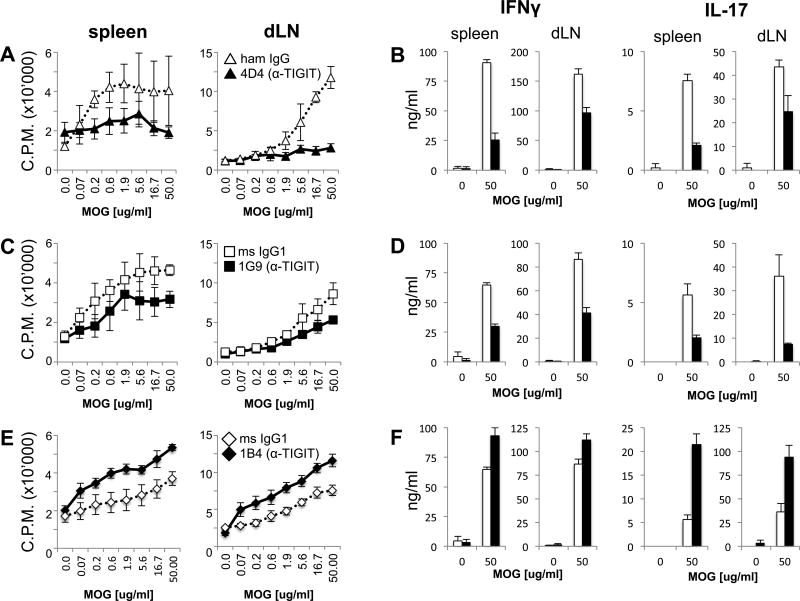
Figure 3. Anti-TIGIT antibodies modulate T cell responses in vivo.
Wild type B6 mice were immunized s.c. with 100 µg MOG35–55 peptide in CFA and received 100 µg anti-TIGIT (or isotype control: Armenian hamster IgG for clone 4D4 or mouse IgG1 for 1G9 and 1B4) antibody i.p on days 0, 2, and 4. On day 10 spleens and lymph nodes were harvested and cells were re-stimulated with MOG35–55 peptide. (dLN: draining lymph node). (A, C, E) After 48 hours, 3H-thymidine was added for the last 18–22 hours before [3H]thymidine incorporation was analyzed to assess proliferation (mean ± SD of triplicate wells from 3–4 animals/group, 3–6 independent experiments). (B, D, F) IFN-γ and IL-17 were measured in the supernatants derived from the same cultures at 48 hours using cytometric bead array. *, P < 0.05; **, P < 0.01; ***, P < 0.001 (Student t test).

