FIGURE 1.
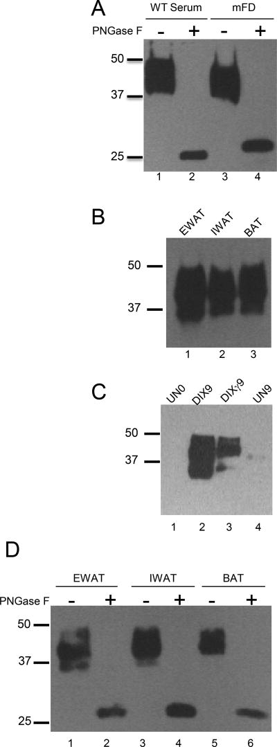
Western blot analysis of mouse FD in serum and adipose tissue. (A) Untreated serum sample from WT mouse (0.3 µl) and also of recombinant mouse FD (30 ng) were treated with PNGase F. (B) Western blot of mouse FD in various adipose tissues: EWAT (Epididymal White Adipose Tissue), IWAT (Inguinal White Adipose Tissue), BAT (Brown Adipose Tissue). (C) Western blot of 1) undifferentiated MEFS prior to differentiation (UN0, lane 1), 2) MEFs differentiated into white adipocytes with the Dexamethasone/Insulin/IBMX protocol (DIX) for 9 d (DIX9, lane 2), 3) MEFs differentiated into brown adipocytes with DIX plus the PPAR-γ agonist troglitazone (DIXγ) protocol for 9 d (DIXγ9, lane 3) and 4) undifferentiated MEFs at the conclusion of the differentiation period (UN9, lane 4). (D) Western blot of mouse FD in various fat tissues (EWAT, IWAT, and BAT) before and after treatment with PNGaseF. These results demonstrate that mouse FD both in serum and adipose tissues is highly and variably N-linked glycosylated.

