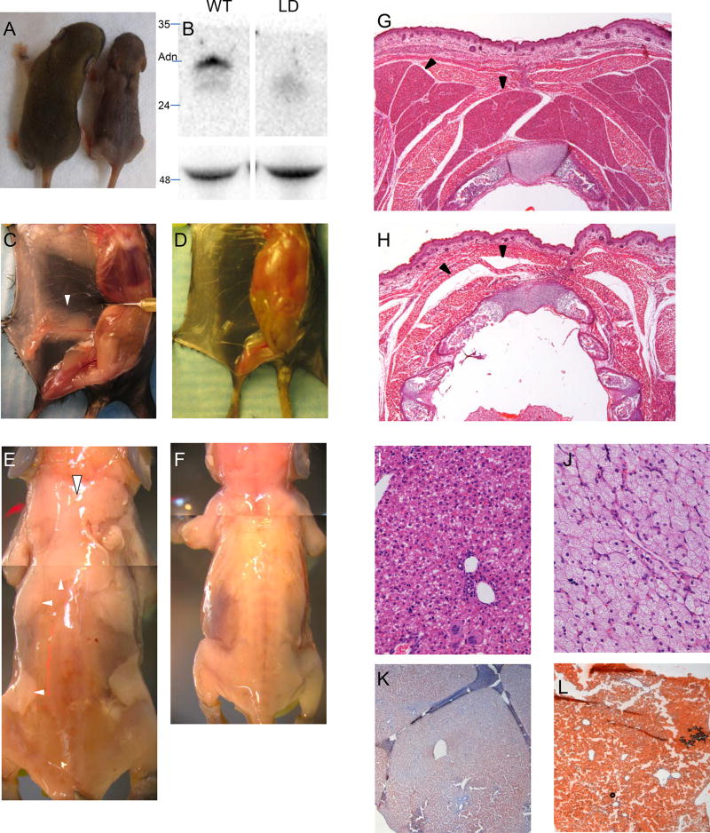
FIGURE 2.
Lipodystrophic mice. (A) WT (left) and LD (right) littermates. Note interscapular defect in LD mouse. (B) Upper, Western blot for adiponectin; lower, loading control (heavy chain). Pinned view of WT (C) and LD (D) mouse showing absence of inguinal fat in LD. (E, F) skinned WT and LD P8 mice showing complete lack of adipose tissue in LD mice. Hematoxylin and Eosin (H&E) staining of newborn postnatal day 0 (P0) WT (G) and LD (H) demonstrating absence of BAT in LD. H&E of liver in WT (I) and LD (J) mice. Oil Red O staining of WT (K) and LD (L) mice. Note the hepatic steatosis in LD mice. White arrowheads, adipose depots in WT mice (missing in LD mice). Black arrowheads, brown adipose in WT mice. In LD mice, black arrowheads point to where BAT should be, but there is only a tissue plane between skeletal muscles.

