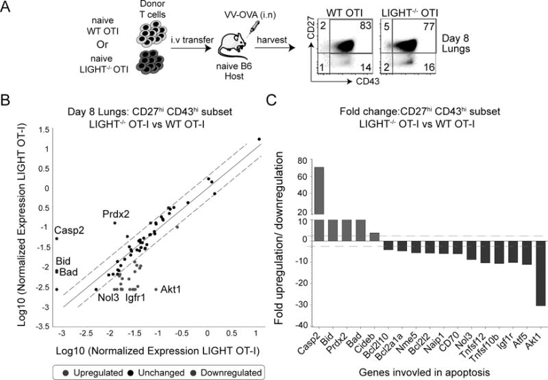
Figure 5. LIGHT regulates the survival of effector CD8 T cells.

(A) Lungs from mice that received WT or LIGHT−/− CD8 T cells were harvested at day 8 post-infection with VacV and stained for CD8, Vα2, Vβ5, CD44, CD27 and CD43. The CD27hiCD43hi subset was sort purified and total mRNA was isolated. (B) Transcript levels of apoptotic genes were measured using affymetrix mouse apoptotic gene arrays and presented as a scatter plot with dotted lines representing 2-fold differences between the two groups. (C) Bars represent fold change in transcript levels of the CD27hiCD43hi subset between LIGHT-deficient CD8 T cells and WT CD8 T cells. Inset: Lung cells from WT and LIGHT−/− OT-I recipient mice were stained intra-nuclearly with pAKT.
