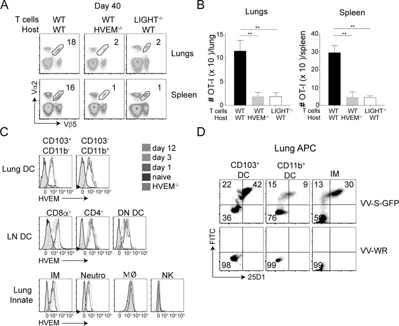
Figure 6. Endogenous HVEM acts as a ligand for LIGHT made by CD8 T cells.

(A & B) Equal numbers (5 × 10⁴) of WT CD8 T cells were adoptively transferred into naive WT and HVEM−/− mice, and compared to LIGHT−/− CD8 T cells transferred into WT mice. All recipient mice were infected with rVacV-WR-OVA (2 × 10⁴ PFU i.n) the following day. At day 40 post-infection, lung and spleen cells were stained for CD8, Vα2, Vβ5, and CD44 and analyzed by flow cytometry. *, P<0.05, **, P<0.01. Similar results were obtained in two independent experiments and results are the mean ± SEM (n = 3 mice/group). (C) Lungs and mediastinal lymph nodes from WT CD8 T cell recipient mice were harvested on day 0 (naïve), 1, 3 and 12 post-infection. Dendritic cells and various innate cells were stained for HVEM. Grey histogram represents cells from HVEM−/− mice. (D) Lung cells were incubated in vitro with either recombinant VV-S-OVA-GFP or VV-WR at MOI of 1. Six-hours post-incubation, cells were stained with antibodies specific for lung DC subsets and inflammatory monocytes and 25D1 that recognizes S-OVA-MHC-I complexes.
