
FIGURE 3.
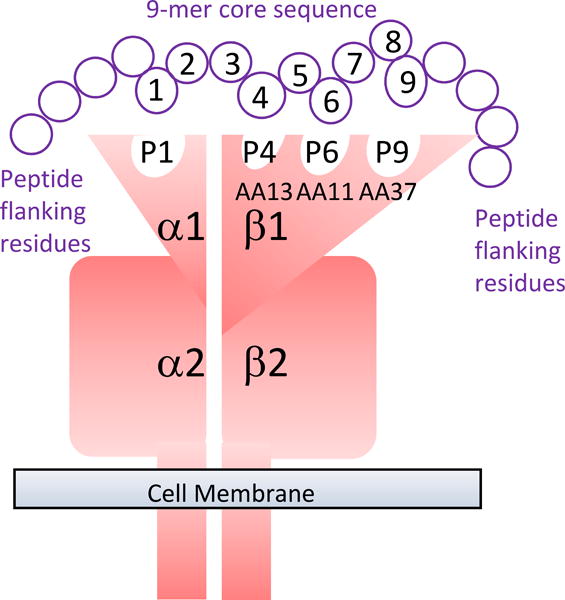
Diagrammatic representation of the DRA/DRB alpha/beta dimer to show how specific amino acids in the binding groove of DRB1 form the pockets that interact with different positions in the 9-mer core epitope of the antigen being presented.
Official websites use .gov
A
.gov website belongs to an official
government organization in the United States.
Secure .gov websites use HTTPS
A lock (
) or https:// means you've safely
connected to the .gov website. Share sensitive
information only on official, secure websites.
Diagrammatic representation of the DRA/DRB alpha/beta dimer to show how specific amino acids in the binding groove of DRB1 form the pockets that interact with different positions in the 9-mer core epitope of the antigen being presented.