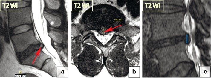
Fig. 13.

Focal disc displacement: extrusion. (a–b) There is an 8-mm focal central L5–S1 extrusion on the sagittal and axial T2-WI. (c) The image shows disc material displacement with complete disruption of the annulus fibrosus; however, the posterior longitudinal ligament remains intact. The posterior aspect of herniation (blue line) is larger than its base (red line) in the sagittal plane, consistent with a full thickness tear of the annulus fibrosus. The herniation material tents the posterior longitudinal ligament without tear. Thus, by definition, this abnormality is a disc extrusion
