
Fig. 23.
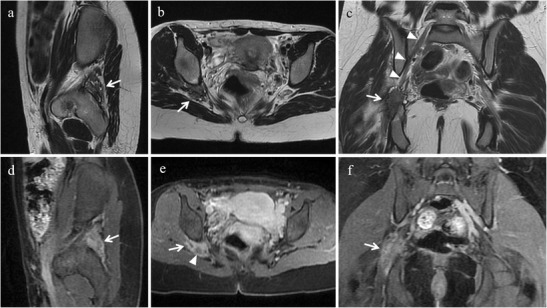
Extrapelvic sciatic nerve endometriosis in a 31-year-old woman who has been suffering from cyclic sciatica for about 2 years. (a) Sagittal, (b) axial and (c) coronal T2-weighted images show hypointense spiculated soft-tissue thickening centred around the right sciatic nerve at the sciatic notch (white arrows). (d) Sagittal, (e) axial and (f) coronal contrast-enhanced fat-suppressed T1-weighted images display enhancement of the mass (white arrows). Note the sciatic nerve cephalad to the lesion (white arrowheads in c) and within the lesion (white arrowhead in e)