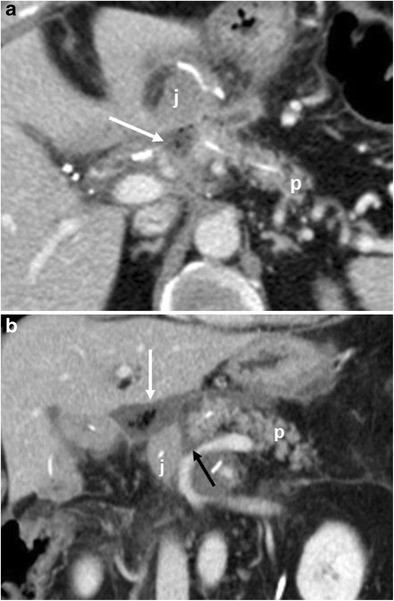
Fig. 14.

Pancreatic fistula. a, b Multiplanar CT images. The pancreatic stump (p) and jejunal loop (j) are visualised. A complete disruption of the pancreatic anastomosis is evident (black arrow in b). At the level of the anastomosis a fluid collection with multiple air bubbles inside is visible (white arrows), a finding that strongly suggests the presence of a pancreatic fistula
