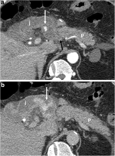
Fig. 18.

a, b Axial CT curvilinear reconstructions show an active extravasation in the arterial phase (white arrow in a) within the lumen of the jejunal anastomotic loop (j). Bleeding becomes more evident in the late phase (white arrow in b). The pancreatic stump is seen (p)
