Fig. 19.

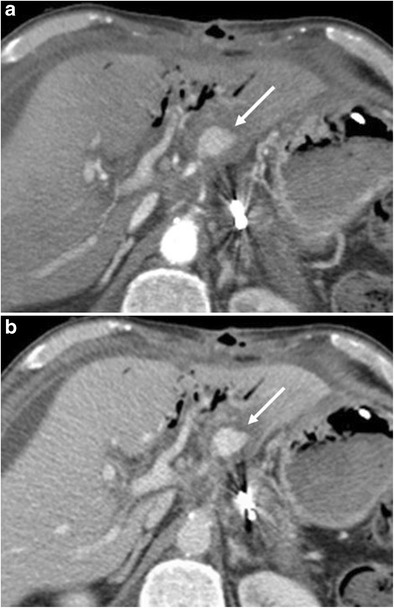
a, b) Axial CT images show an active extravasation in the arterial phase (white arrow in a) coming from the common hepatic artery after a Whipple procedure. Bleeding becomes more evident in the venous phase (white arrow in b)

a, b) Axial CT images show an active extravasation in the arterial phase (white arrow in a) coming from the common hepatic artery after a Whipple procedure. Bleeding becomes more evident in the venous phase (white arrow in b)