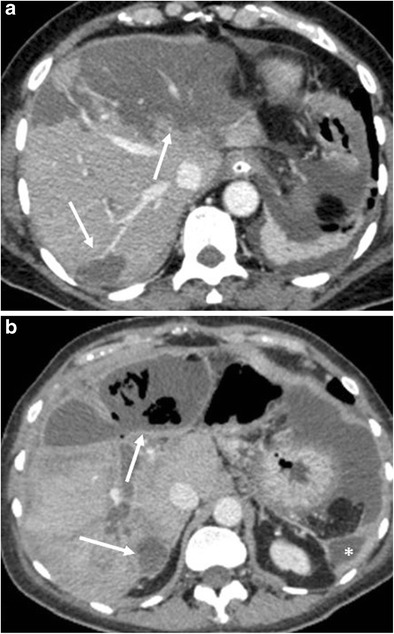
Fig. 20.

Hepatic infarction. a CT image shows multiple hypodense and hypovascular areas of infarction (white arrows) following a pancreaticoduodenectomy. b CT image 4 weeks later in the same patient. The areas of infarction show a thickened and enhancing wall with multiple air bubbles within (white arrows), findings consistent with hepatic abscesses. Another abscess is visible at the level of the left lateroconal fascia (*)
