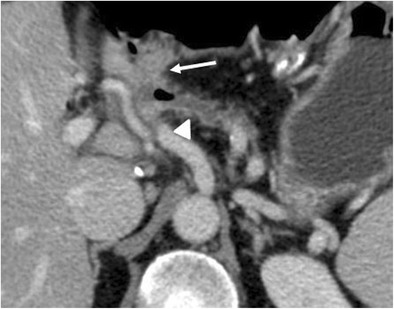
Fig. 26.

Anastomotic stricture. Axial CT image shows a dilation of the main pancreatic duct associated with atrophy of the pancreatic parenchyma (white arrowhead). No signs of local tumour recurrence are seen at the pancreatic anastomosis (white arrow)
