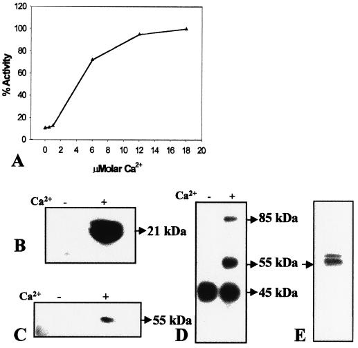
Figure 3.
CDPK activity and protein in soluble protein extract of torpedo-stage somatic embryos. A, Effect of micromolar Ca2+ on the phosphorylation of histone III-S. One-hundred percent activity represents a specific activity of 0.48 pmol mg−1 min−1. B, Autoradiogram of an SDS-polyacrylamide gel showing the position of histone III-S phosphorylated in the presence of 0.2 mm (+) or absence (−) of Ca2+ by incubation with protein extracts (10 μg) prior to electrophoresis. C, In-gel histone III-S phosphorylation assay in which 50 μg of protein extract was loaded in each lane. Band on the autoradiogram indicates the position of histone kinase activity on the gel. D, In vitro CDPK assay in the absence of an exogenous substrate followed by separation on 10% (w/v) SDS-polyacrylamide gel. Bands at 55 and 85 kD on the autoradiogram indicate positions of proteins that are phosphorylated in presence of 0.2 mm Ca2+. E, Western-blot analysis of 50 μg of protein using polyclonal anti-soybean CDPK.

