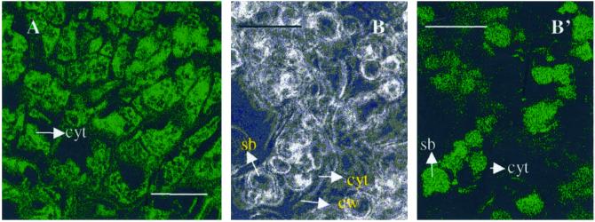
Figure 9.
Immunofluorescence localization of swCDPK. A, Cytoplasmic localization of swCDPK in cells of embryos. B, Bright-field image showing cells of endosperm of the G2 stage of germination; B′, immunofluorescence localization of swCDPK in storage bodies of the endosperm cells. cyt, Cytoplasm; cw, cell wall; sb; storage bodies. Scale bars = 30 μm.

