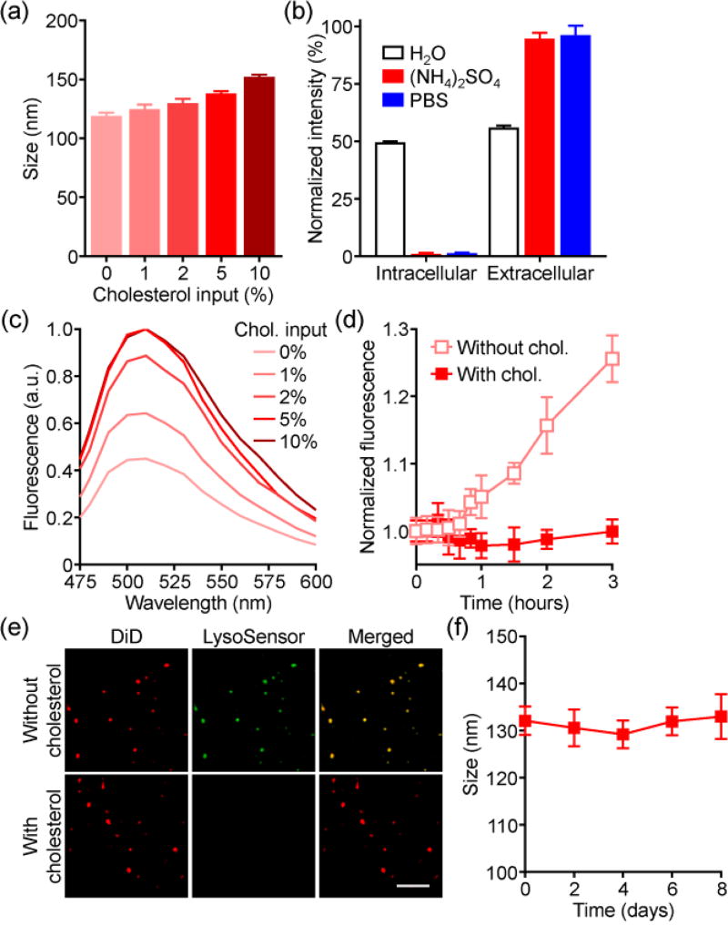
Figure 2.
Characterization of cholesterol-enriched RBC vesicles. a) Size of vesicles with varying cholesterol inputs (n = 3; mean ± SD). b) Dot blot intensities of various RBC vesicles probed with antibodies against the intracellular or extracellular domains of CD47 (n = 3; mean ± SD). c) Fluorescence spectrum of ammonium sulfate-loaded RBC vesicles when co-loaded with a pH-sensitive LysoSensor Green dye. d) Leakage of calcein from RBC vesicles (n = 3; mean ± SD). e) Confocal fluorescence imaging of ammonium sulfate-loaded RBC vesicles co-loaded with LysoSensor Green (red: DiD, green: LysoSensor Green; scale bar = 5 µm). f) Size of enriched RBC vesicles over time in serum-containing media (n = 3; mean ± SD).

