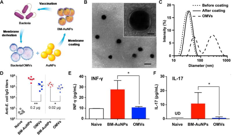
Figure 2.
Membrane-coated nanoparticles for antibacterial vaccination. (A) Schematic illustration of bacterial membrane-coated nanoparticles for modulating antibacterial immunity. Outer membrane vesicles (OMVs) are collected from source bacteria and coated onto gold nanoparticles (AuNPs) to form bacterial membrane-coated AuNPs (BM-AuNPs). When used for vaccination, BM-AuNPs can elicit specific immunity against the source bacteria. (B) A representative electron microscopy image showing the core-shell structure of the BM-AuNPs negatively stained with uranyl acetate (scale bar, 50 nm). Inset: a zoomed-in view of a single BM-AuNP (scale bar, 10 nm). (C) Size intensity distribution of OMVs and AuNPs, before and after coating with bacterial membrane, as measured by dynamic light scattering. (D) BM-AuNPs elicit strong anti-E. coli IgG titers 21 days after vaccination. (E, F) BM-AuNPs induce pronounced bacterium-specific T cell activation in vivo. On day 21 after vaccination, splenocytes were collected and stimulated with E. coli bacteria. After 72 hours of culture, the levels of (E) IFN-γ and (F) IL-17 were quantified by ELISA. Reprinted with permission from ref 26. Copyright 2015 American Chemical Society.

