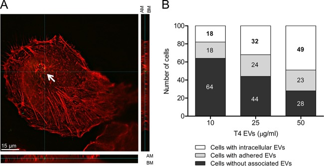
FIG 2 .
EVs are internalized by A549 lung epithelial cells. (A) High-resolution immunofluorescence microscopy imaging of A549 cells incubated with EVs (50 μg/ml) for 6 h and stained with phalloidin, used as an epithelial cell marker (red), and pneumolysin, used as an EV marker (green). In this representative image, the complete thickness of epithelial cells has been imaged in 21 z-stacks. The internalized vesicle, indicated with a white arrow, has been captured in z-stack 11, which is displayed from the top (A) and in XZ (A [bottom]) in YZ (A [right]) orthogonal views to demonstrate intracellular localization. AM, apical membrane; BM, basolateral membrane. (B) Quantification of internalization of vesicles in A549 cells treated with 10, 25, and 50 µg/ml of EVs from T4. For each concentration, 100 cells were analyzed and the number of cells with intracellular or extracellular signal or of cells without EV-associated signal is shown.

