
Figure 1.
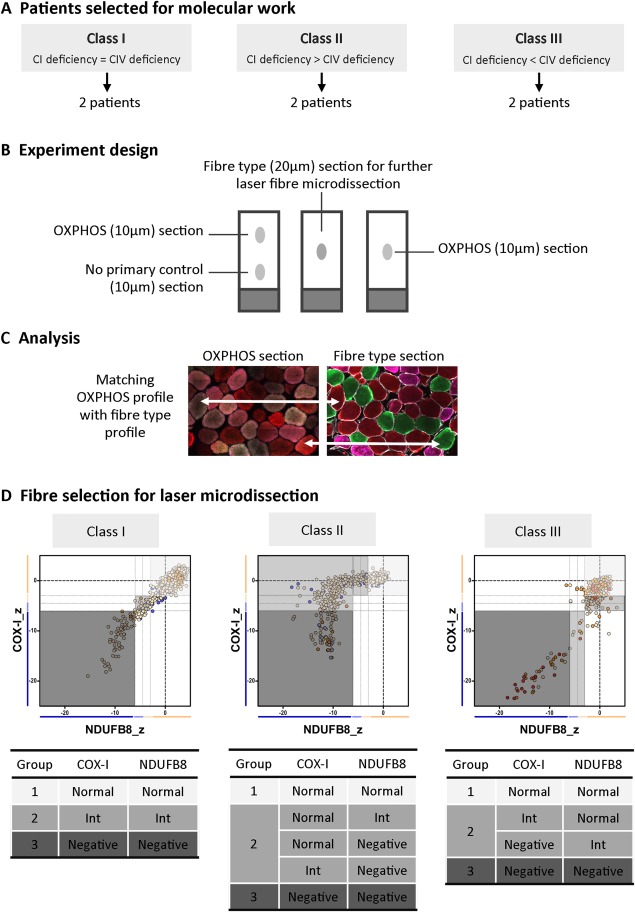
Design of the molecular study. (A) Patients were first classified into different classes (I–III) according to their mitochondrial respiratory chain profile, and then 2 patients from each class were selected for single fiber analysis based on their biopsy size. (B) Four serial muscle sections were taken from each patient's biopsies for oxidative phosphorylation (OXPHOS) and fiber type analysis. (C) The OXPHOS section was sequentially incubated with all primary and secondary antibodies (green = COX‐I, red = porin, purple= NDUFB8), whereas the no primary control section was inbubated only with laminin antibody and all secondary antibodies. The fiber type section was incubated with antibodies detecting type I (green) and type II (red = IIa, purple = IIx) myosin heavy chain. The OXPHOS and fiber type serial sections were manually overlaid to match fibers across sections. (D) Fibers were then selected for microdissection, with careful consideration of the OXPHOS group (1–3) and fiber type. CI = complex I; CIV = complex IV; Int = intermediate.