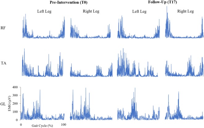
Figure 2.
Bilateral, rectified electromyogram (EMG) traces of the rectus femoris (RF), tibialis anterior (TA), and gastrocnemius lateralis (GL) at pre-intervention and follow-up for a representative participant with Parkinson’s disease. Y-axes of the EMG traces are scaled consistently across each muscle.

