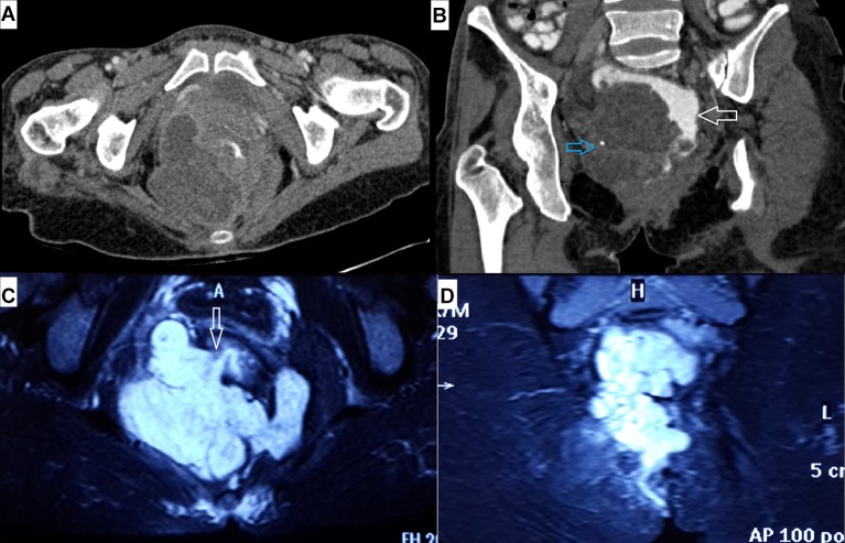
Figure 1.
(A) Axial and (B) oblique coronal contrast-enhanced CT images showing large hypodense pelvic mass impinging on rectum (white arrow in B), displacing it towards left. Also noted a tiny flake of calcification within the mass (blue arrow in B). Fat-suppressed MRI T2-weighted images in (C) axial and (D) coronal plane showing large lobulated T2 hyperintense mass involving both ischiorectal fossae mimicking horseshoe abscess. The mass is in relation to the tract of the fistula, and tumour-free internal opening is communicating with anal canal at 8 o’clock position (arrow in C).

