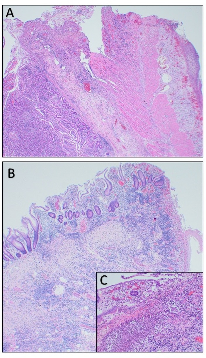
Figure 2.
A shows acute transmural ischaemic necrosis of the small bowel with perforation and peritonitis (H&E magnification ×40). B shows acute ischaemic changes to the small bowel with ulceration (H&E magnification ×40). C is a higher power image of acute ischaemic changes to the small bowel mucosa with ulceration (H&E magnification ×250).

