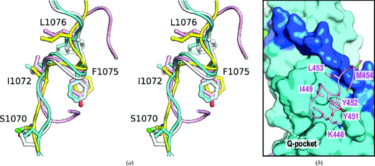
Figure 4.
(a) Stereoview of the structural overlay of ZRANB3 APIM (yellow) and PIPMs: p21 (PDB entry 1axc; silver), Pol-ι (PDB entry 2zvm; pale pink) and Pol-η (PDB entry 2zvk; pale cyan; Hishiki et al., 2009 ▸). Residues of ZRANB3 APIM are labelled. (b) Binding of the noncanonical PIPM of Pol-ι to PCNA. The structure of Pol-ι bound to PCNA is shown as a pale pink tube (PDB entry 2zvm). Residues of the ‘hydrophobic plug’ (Ile449, Tyr452 and Leu453) are shown in stick representation and are labelled. Residues engaged in intramolecular contacts (Lys446 and Tyr451) are also shown. PCNA is shown as a surface representation and the colours correspond to those in the left panel of Fig. 2 ▸(c).

