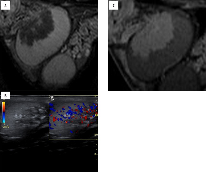
Figure 1.
(Case 4) Asymptomatic, 23-year-old boy with CAH, ultrasound performed 2 years before was normal. Ultrasonography (A) shows a hypervascular heterogeneous lesion located around the mediastinum testis. Also, coarse calcifications and fibrous septation are seen. Sagittal (B) T2-weighted image shows an intra-testicular mass that is hypointense, as compared with normal testicular tissue. Sagittal postcontrast (C) T1-weighted image reveals a marked enhancement that is greater than that of normal parenchyma.

