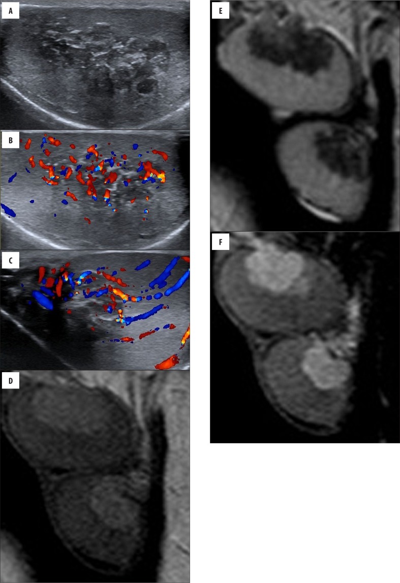
Figure 2.
(Case 9) Bilateral palpable testicular masses in a 13-year-old boy with CAH. Longitudinal (A, B) gray-scale and color Doppler sonogram images of the left testicle show hypervascular heterogeneous hypoechoic lesions within mediastinum testis. Color Doppler US (C) of the right testicle shows normal testicular vessels coursing undisturbed through the mass; the vessels are not displaced and have a normal caliber. Sagittal (D) T2-weighted MRI shows intra-testicular masses that are hypointense, as compared with normal testicular tissue. Sagittal (E) pre-contrast T1-weighted image shows bilateral testicular lesions that are slightly hyperintense. Sagittal (F) postcontrast T1-weighted image reveals marked enhancement in the masses that is greater than that of normal parenchyma.

