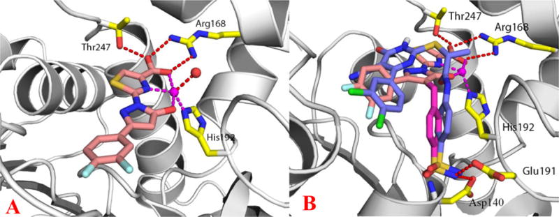
Figure 2.

(A) Crystal structure of inhibitor 27 bound to LDHA in complex with zinc. The inhibitor is shown in sticks with salmon-colored carbons. The protein is shown in ribbon representation and the metal zinc is shown as a magenta sphere. A water molecule (red sphere) and protein residues R168, H192 and T247 (yellow-colored carbons) are coordinated with Zn or form H-bonding interactions with the inhibitor. PDB: 5W8I (B) Inhibitor 33 docked in the binding pocket of LDHA and overlaid with 4 (purple-colored carbons). The benzyl sulfonamide moiety shown as magenta-colored carbons.
