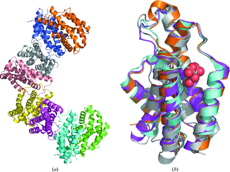
Figure 1.
(a) Ribbon diagram of the tetramer of dimers for chorismate mutase from B. phymatum. (b) Superposed monomers of chorismate mutases with AroQγ topology from B. phymatum (PDB entry 5ts9; magenta), B. thailandensis (PDB entry 4oj7; cyan), M. tuberculosis (PDB entry 2fp2; gold) and Y. pestis (PDB entry 2gbb; gray with the citrate molecule shown as spheres).

