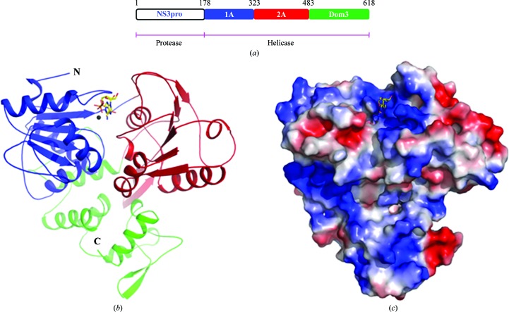
Figure 1.
Overall structure of the helicase from ZIKV. (a) Diagrammatic representation of the domain boundaries of the NS3 protein from ZIKV. Crystal structures of the region encompassing residues 178–618 are reported in this study. (b) Cartoon representation of the structure of the helicase from ZIKV bound to AMPPNP and Mn2+. The convention used for colouring the domains of the structure is shown in (a). AMPPNP is shown as sticks, while the Mn2+ ion is shown as a sphere. The N- and C-termini of the protein are marked. (c) Surface electrostatic potential representation of the structure of the helicase from ZIKV. Blue represents positive potential and red negative potential.

