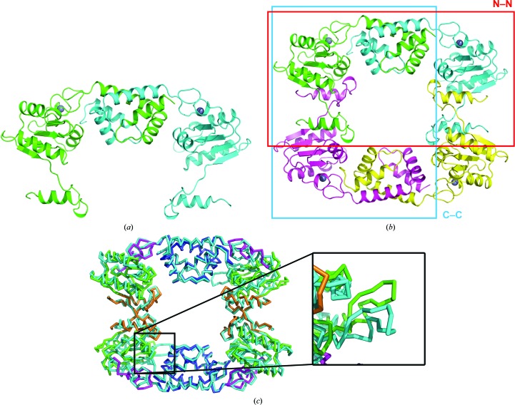
Figure 3.
The PaRecR dimer and tetramer structures. (a) The PaRecR N–N dimer structure, shown in ribbon representation. Subunit A is coloured green and subunit B is coloured cyan. (b) The PaRecR tetramer structure shown in ribbon representation. Subunit A is coloured green, subunit B is coloured cyan, subunit A′ is coloured magenta and subunit B′ is coloured yellow. The N–N and C–C dimers are shown by red and blue boxes, respectively. (c) Superposition of PaRecR and T. tengcongensis RecR tetramers. Both tetramers are shown in PyMOL ribbon representation; PaRecR is coloured according to structural domain (blue, HhH motif; magenta, zinc-finger motif; green, Toprim domain; orange, Walker B motif) and T. tengcongensis RecR is coloured cyan. Inset: comparison of loop 106–121 of the two structures.

