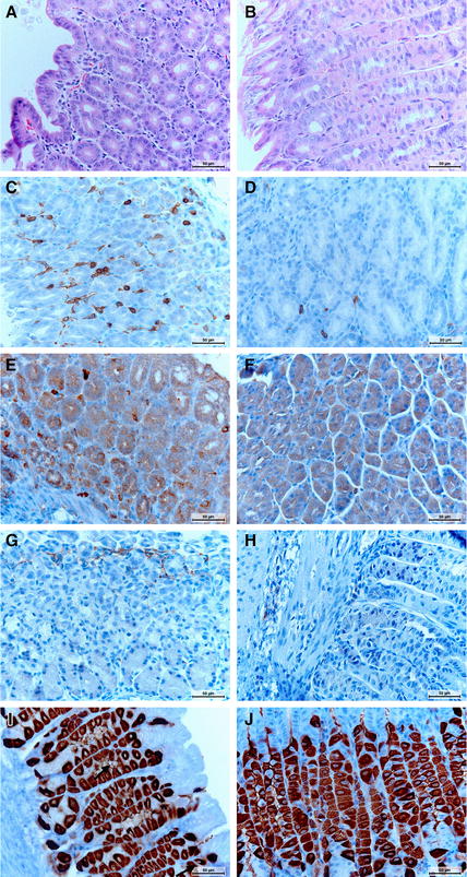
Figure 2.

Microscopic visualization of immune cells, apoptotic cells, proliferative cells and parietal cells in the mouse stomach. A, B HE-staining showing infiltration with mononuclear and polymorphonuclear cells in H. suis infected mice without (A) and with bambermycin (B) supplementation. The infiltration is less severe during bambermycin supplementation. Original magnification: ×400. C, D CD3-staining showing infiltration with T-cells (brown) in H. suis infected mice without (C) and with bambermycin (D) supplementation. The infiltration is less severe during bambermycin supplementation. Original magnification: ×400. E, F CD20-staining showing infiltration with B-cells (dark brown) in mice infected with (E) and without H. suis (F). The infiltration is more severe during H. suis infection. Original magnification: ×400. G, H F4/80 staining showing infiltration with macrophages (brown) in mice infected with (G) and without (H) H. suis. The infiltration is more severe during H. suis infection. Original magnification: ×400. I, J H+/K+ATPase-staining showing parietal cells (brown) in mice infected with (I) and without (J) H. suis. The number of parietal cells is lower during H. suis infection. H. suis bacteria can be seen in the lumen of the gastric glands (arrow). Original magnification: ×400.
