Fig. 1.

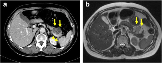
Findings from preoperative CT and MRI. CT and MRI show a cystic neoplasm (arrow) in the pancreatic body. Cyst diameter is 35 mm. a CT. b T2-weighted MRI. Splenic vein was normal status (arrowhead)

Findings from preoperative CT and MRI. CT and MRI show a cystic neoplasm (arrow) in the pancreatic body. Cyst diameter is 35 mm. a CT. b T2-weighted MRI. Splenic vein was normal status (arrowhead)