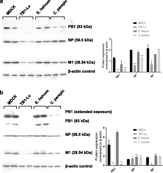
Fig. 5.

Variation in viral protein production between infected bat cells. All three species of bat cells and MDCK cells were infected with human USSR H1N1 (a) or avian H2N3 (b) virus at 0.5 MOI for 24 h for the detection of viral PB1, NP and M1 along with detection of β-actin as loading control. Differential viral protein expression of PB1, NP and M1 was evident between the three bat cell types for each virus (a and b). All three viral proteins were strongly expressed in MDCK and E. helvum cells with each virus. USSR H1N1 virus infected TB1-lu cells showed weak expression of each viral protein; similarly avian H2N3 virus infected TB1-Lu cells showed faint presence of PB1 and M1 proteins. Infected C. perspic cells displayed an intermediate pattern of viral protein expression that was between that of correspondingly infected E. helvum and TB1-Lu cells. Insets are corresponding densitometric quantification of Western blotting results. Significance indicated is in relation to corresponding MDCK cells
