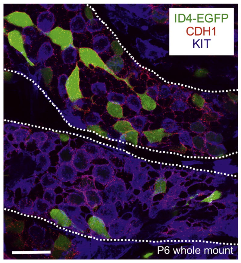
Fig. 5.

Whole-mount immunostaining of P6 Id4-eGfp testis cords. Maximum intensity Z-stack projection of isolated testis cords from transgenic Id4-eGfp P6 mice (ID4-EGFP epifluorescence in green). Antibody staining was performed for the undifferentiated marker CDH1 (in red) and the differentiating marker KIT (in blue). Cords are outlined with white dashed lines. Scale bar = 25 μm.
