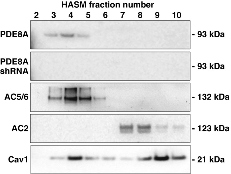
Figure 6.
Western blot analysis of PDE8A in fractions derived from sucrose density centrifugation of control HASM cells or cells incubated 72 hours with recombinant lentivirus-expressing PDE8A shRNA. Cells were fractionated using a nondetergent method and separated by sucrose density centrifugation (see Methods section of main text). Gradients were collected in ten 0.5-ml fractions, separated by SDS-PAGE, and analyzed by immunoblotting using a primary antibody for PDE8A. Fractions were also probed for native expression of AC6, AC2, and caveolin-1 (Cav1) using antibodies for AC5/6, AC2, and Cav1 to show appropriate fractionation. Both control and PDE8A-knockdown HASM cells displayed similar distributions of Cav1, AC5/6, and AC2. Fractions 3–5 contain buoyant lipid raft membranes, whereas fractions 6–10 contain the rest of the cellular material. Shown are regions of the gels at the approximate molecular weight of the expected immunoreactive band. Images shown are representative of three experiments.

