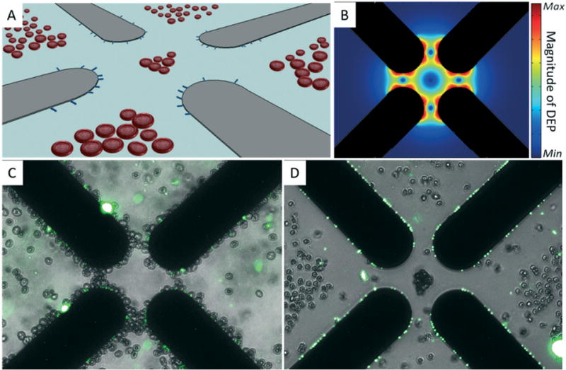
Fig. 2.

Discrimination of bacteria and human blood cells via chemically mediated dielectrophoresis. (A) Illustration of co-planar quadrupole microelectrodes (gray objects) used to separate bacteria (blue rods) from blood cells by DEP. Blood cells are repelled away from electric field maxima located at the electrode edges while bacteria are pulled toward these regions. (B) Simulated magnitude of the DEP force near the quadrupole micro-electrode tips (black shadows). (C) Merged microphotographs of E. coli labeled with the fluorescent dye SYTO9 (green channel) and erythrocytes (bright-field channel) suspended in 40 mS m−1 280 μM D-mannitol. Both cell types were collected by pDEP at microelectrode edges using a sinusoidal signal of 20 MHz and 4 Vpp. (D) Merged microphotographs of labeled E. coli (green channel) and erythrocytes (bright-field channel) suspended in 40 mS m−1 280 μM D-mannitol and 260 nM monensin. Bacteria and blood cells were separated using the same DEP signal as in (C).
