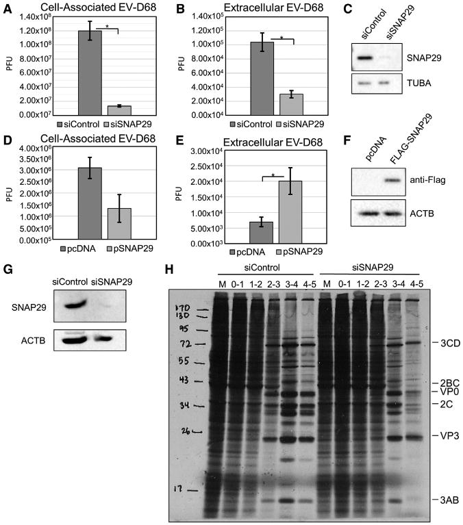
Figure 6. SNAP29 Affects EV-D68 Infection in H1HeLa Cells.

(A and B) Cells were transfected with a siRNA targeting SNAP29 40 hr prior to infection and then infected with EV-D68 at an MOI of 0.1 for 5 hr. Viral titers were analyzed by plaque assay for both cell-associated virus (A; p = 0.018) and extracellular virus (B; p = 0.012).
(C) Parallel samples were analyzed by western blot for SNAP29. H1HeLa cells were transfected with a plasmid containing FLAG-SNAP29 40 hr prior to infection.
(D and E)Transfected H1HeLa cells were infected at an MOI of 0.1 with EV-D68 for 5 hr. Viral titers were analyzed by plaque assay for both cell-associated virus (D) and extracellular virus (E; p = 0.017).
(F) Western blot to examine FLAG-SNAP29 ectopic expression. Western blots are representative of 3 independent experiments.
(G and H) H1HeLa cells were transfected with siSNAP29 40 hr prior to infection, infected at an MOI of 25 with EV-D68, and metabolically labeled each hour with 35-S methionine.
(G) Samples were analyzed for SNAP29 in knockdown cells.
(H) Samples were subjected to SDS-PAGE and exposed to X-ray film.
Viral titers are represented as the mean ± SEM. Statistical tests were done using Student's t test with statistical significance set at *p ≤ 0.05.
