
Fig. 1.
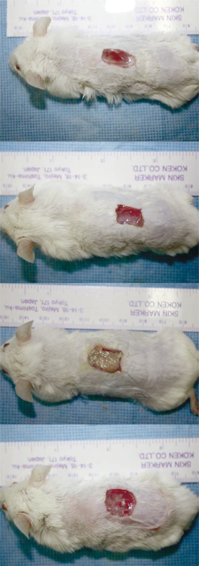
Gross findings of the full-thickness wound on the dorsum of the mouse on days 10: the Oncorhynchus mykiss-derived polydeoxyribonucleotide (PDRN) injection group, the Oncorhynchus keta-derived PDRN injection group, the O. keta-derived PDRN cream group, the normal saline soaked dressing group, respectively. There was no delayed wound healing response in all the groups across all time periods. In the O. keta-derived PDRN cream group, the wound appeared as if it had impurities and was less clean when compared with the other groups. However, when the cream was removed, wound healing could be seen from inside of the wound.