Fig. 3.

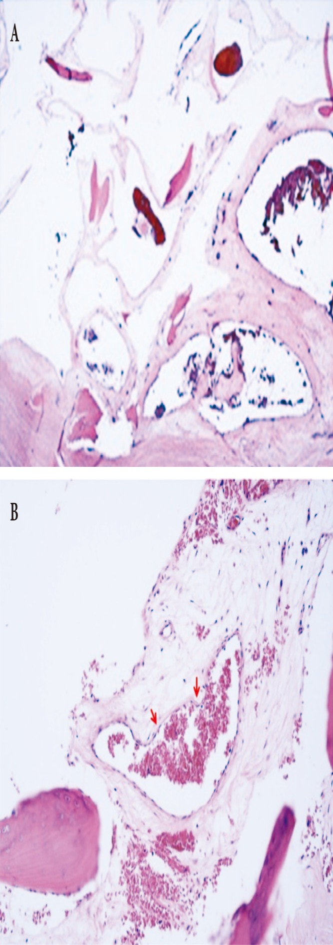
Histopathologic findings. (A) In a low-power view, multiple dilated vascular spaces are present between pre-existing bony trabeculae (H&E, ×40). (B) These vascular spaces are lined by flattened endothelial cells (arrows) (H&E, ×200).

Histopathologic findings. (A) In a low-power view, multiple dilated vascular spaces are present between pre-existing bony trabeculae (H&E, ×40). (B) These vascular spaces are lined by flattened endothelial cells (arrows) (H&E, ×200).