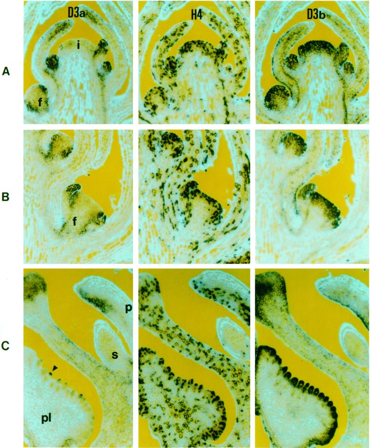
Figure 5.
RNA in situ hybridization of consecutive longitudinal sections of inflorescence apices with RNA probes from the cyclin D3a (D3a), histone H4 (H4), and cyclin D3b (D3b) genes. In A through C, RNA is indicated by the dark-blue color, while epifluorescence was used to reveal calcofluor-stained (light blue) cell walls. A, Inflorescence apex containing the inflorescence meristem (i) and floral meristems at three different stages (f). B, Sections from a florotypic floral meristem (f). C, Sections through an unopened floral bud showing petal (p), staminode (s), carpel wall (c), and placenta (pl).

