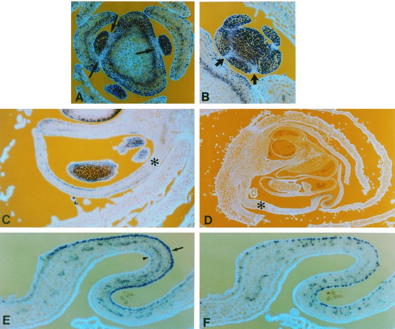
Figure 6.
Patterns of cyclin D3b expression during floral development. Sections through young floral meristems at about node 7 (A) and 14 (B) showing regions of reduced cyclin D3b expression (arrows) surrounding the young meristems (A) and sepal primordia (B). Sections through floral buds before (C) and during (D) the initiation of petal folding, probed with cyclin D3b gene. Ventral petals are indicated with an asterisk. E and F, Near consecutive sections of folded regions of the ventral petal shown in D probed with cyclin D3b (E) or histone H4 (F); the inner surface of the petal is indicated by an arrow and the outer surface by an arrowhead.

