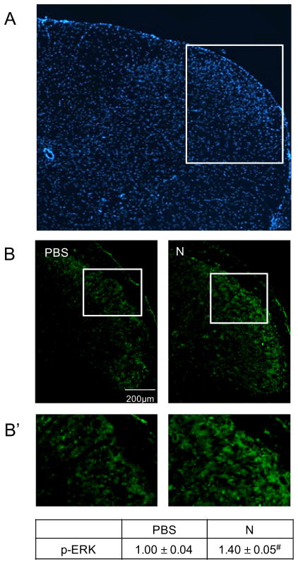
Fig. 2.
Nicotine administration resulted in an increased p-ERK expression in the upper spinal cord tissue containing the spinal trigeminal nucleus. (A) Representative image of the upper spinal cord tissue stained with the nuclear dye DAPI. (B) Images acquired at 100× magnification representing spinal cord sections taken from vehicle control (PBS) and nicotine (N)-treated animals immunostained for p-ERK. (B′) Enlarged image of medullary horn delineated in panel B. The change in p-ERK staining intensities is reported as the average fold change±SEM as compared to mean levels in control samples that was set equal to 1 (n=3). #P ≤ 0.01 when compared to control values.

